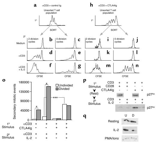
Figure 2.
B7-mediated costimulation and cell division differentially regulate secondary T cell proliferation. (a–o) CFSE-labeled spleen and lymph node cells were stimulated with anti-CD3 in the presence of human IgG (a–g; first and second sets of columns in o) or CTLA4Ig (h–n; third and fourth sets of columns in o). Cultures were rested for 48 hours, and Thy1.2+ cells that had divided twice (b, d, f, i, k, and m; shaded bars in o) or had remained undivided (c, e, g, j, l, and n; open bars in o) following primary stimulation were purified by FACS. The sorted T cells were cultured with irradiated APCs and restimulated with anti-CD3 in the presence or absence of exogenous IL-2; proliferation was assessed 4 days later by flow cytometry. One representative experiment for each condition (a–g and h–n) is depicted graphically. The mean secondary mitotic events of separate experiments (n = 2 [first and second sets of columns] or n = 4 [third and fourth sets of columns]) are plotted in o. Statistically significant differences were assessed by paired t test and are denoted by brackets: *P < 0.05; ***P < 0.001. (p) Lymph node and spleen cells were cultured with anti-CD3 in combination with anti-CD28 antibody (first and second lanes) or CTLA4Ig (third and fourth lanes). The cultures were rested for 24 hours, and the T cells were restimulated with anti-CD3–coated beads for 48 hours in the presence (second and fourth lanes) or absence (first and third lanes) of IL-2. Live cells were harvested after the primary stimulus (top panels) and after the secondary stimulus (bottom panel) by isolation over Ficoll, and lysates were subjected to immunoblot analysis using antibodies against p27kip1 (top and bottom panels) or actin (data not shown). The results shown are representative of two independent experiments. (q) Primary, CFSE-labeled T cells were primed with anti-CD3 as in p and rested (top panel), and a portion of the cells were restimulated with either 50 U/ml IL-2 for 48 hours (middle panel) or PMA/ionomycin (PMA/Iono) for 24 hours (bottom panel). The live, CD4+ cells were then sorted into fractions that had divided two or more times (right lane, “D”), or had remained undivided during the culture period (left lane, “U”). The cells were lysed, and equal cell equivalents were assessed for p27kip1 content by immunoblot analysis. The data shown are representative of two independent experiments.

