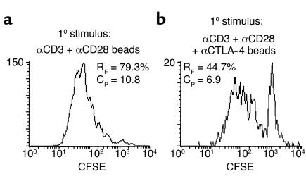
Figure 3.
Regulation of primary T cell division by B7–CTLA-4 interactions. (a) Pooled BALB/c lymph node and spleen cells were labeled with CFSE, and T cells were subjected to co-crosslinking of TCR and CD28 by the addition of polystyrene beads coated with anti-CD3 (1 μg/ml), anti-CD28 (1 μg/ml), and control hamster IgG (1 μg/ml). (b) In separate cultures, T cells were subjected to co-crosslinking of TCR, CD28, and CTLA-4 by the addition of polystyrene beads coated with anti-CD3 (1 μg/ml), anti-CD28 (1 μg/ml), and anti–CTLA-4 (1 μg/ml). Proliferation of the CD4+ T cell subset was assessed by flow cytometry 3 days later. The frequency of precursor T cells that divided in response to stimulus (RF), and the number of daughter T cells generated by the average responding precursor T cell (CP), were calculated as described previously (2, 3). The data are representative of two separate experiments.

