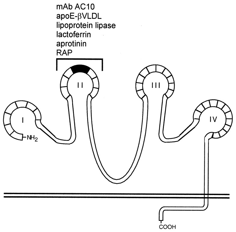
Figure 6.
Ligand binding map of megalin. Megalin contains four clusters (I–IV) of ligand-binding repeats that most likely attain a complex tertiary structure stabilized by numerous intramolecular disulfide bonds. Each of the four clusters are comprised of a specific number of homologous cysteine-rich repeats (boxed areas). In analogy to the LDL receptor, each cluster represents a putative ligand binding site containing both acidic and basic amino acid residues that may facilitate high-affinity, charge-dependent receptor–ligand interactions. The second cluster of ligand-binding repeats (bracket) contains a binding site (amino acids 1111–1210, shaded area) for apoE-βVLDL, lactoferrin, aprotinin, lipoprotein lipase, and RAP. This region also serves as a binding site for circulating autoimmune antibodies found in Heymann nephritis (29).

