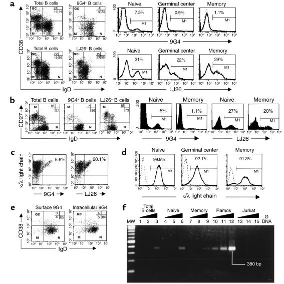
Figure 1.
Representation of VH4-34 cells in human B cell subsets. (a) Distribution of total, VH4-34 (9G4) and VH3 (LJ26) B cells into naive (N), germinal center (GC), and memory (M) subsets in a representative tonsil. Histograms demonstrate the frequency of 9G4 and LJ26 cells in each subset. Bold lines represent VH4-34 and VH3 cells. Isotype antibody controls for 9G4 are shown as superimposed dashed lines. (b) Similar Vresults were obtained with peripheral blood B cells. (c) 9G4 and LJ26 cells express similar levels of sIg (determined with anti-κ/λ antibodies). (d) Analysis of the sIg expression by tonsil B cells. At least 90% of cells in each subset express detectable levels of sIg. (e) Tonsil B cells were permeabilized and analyzed for either surface or cytoplasmic expression of VH4-34. (f) PCR analysis of VH4-34 rearrangements. Samples are as follows: MW, 100 bp ladder; Ø DNA, negative control without input DNA; 1–3, total tonsil B cells; 4–6, naive B cells; 7–9, memory B cells; 10–12, VH4-34–expressing Ramos B cell lymphoma; 13–15, Jurkat T cells. The following amounts of DNA template were used: lanes 1, 4, 7, 10, and 13 (33 ng); lanes 2, 5, 8, 11, and 14 (100 ng); lanes 3, 6, 9, 12, and 15 (300 ng). Noticeable amplification of rearranged VH4-34 was detected in memory cells only after 35 PCR cycles with the highest amount of template DNA (300 ng). The amount of product obtained with memory cells was four to ten times lower than that obtained with naive B cells (average of four experiments). Calculations based on equivalent amounts of Ramos cells or Ramos DNA indicate that less than 2% of memory cells express VH4-34.

