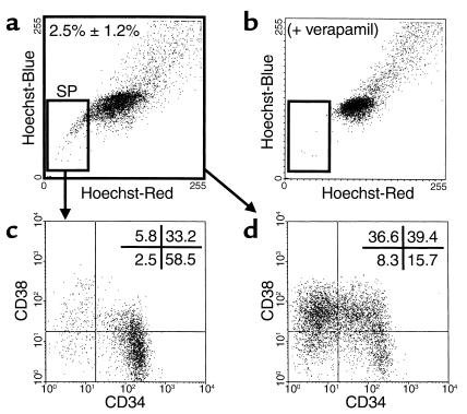
Figure 1.
Representative FACS dot plot showing the presence and phenotypes of SP cells in the lin– fraction of human fetal liver cells. Cells were depleted of lin+ cells and stained with Hoechst 33342 and antibodies to CD34 and CD38 as described in Methods. (a) Small gated cell population identifies the SP cells (2.5% ± 1.2% of the total lin– fetal liver population; n = 9) that disappear in the presence of verapamil (b). The distribution of cells according to their expression of CD34 and CD38 in the SP (c) and total (d) fractions of the lin– fetal liver population analyzed here is also shown.

