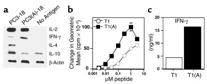
Figure 2.
Higher-affinity binding peptide T1(A) skews T cells toward a Th1 response. (a) Inguinal and periaortic lymph node cells were harvested from animals immunized with PCLUS 3-18IIIB (PC3-18) or PCLUS 3(A)-18IIIB [PC3(A)-18] and stimulated overnight with the immunizing peptide at 3 μM. Total RNA was isolated from positively selected CD4+ T cells 20 hours after in vitro stimulation and semiquantitative RT-PCR performed under nonsaturating conditions to compare relative cytokine mRNA levels. These results were repeated with similar results in two additional experiments. (b) Stimulation of a T1 CD4+ T cell line with peptide T1(A) elicited greater proliferation than when stimulated with T1. A similar level of proliferation was obtained at a 30-fold lower concentration of peptide T1(A); compare proliferation at 0.1μM T1(A) versus 3μM T1. (c) IFN-γ production over 48 hours as measured by ELISA from a T1-specific CD4+ T cell clone stimulated with peptide T1 or T1(A).

