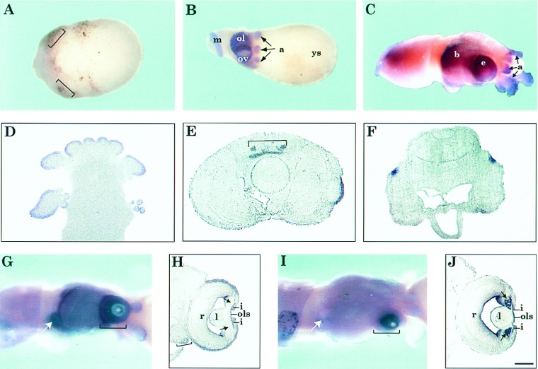
Figure 3.
Expression of Pax-6 during L. opalescens embryonic development. (A–C, G, and I) Whole mount in situ hybridization of squid embryos using Pax-6 (A–C and G) and S-crystallin (I) antisense riboprobes. Sense probes were used as controls (data not shown). (D–F, H, and J) Frontal, 10-μm plastic sections of embryos hybridized as whole mount with Pax-6 (D–F and H) and S-crystallin (J) antisense probes (20). (A) Stage 17 embryo. Brackets indicate the eye primordia where first traces of Pax-6 expression are detected. (B) Stage 20 embryo. Arrows point to the arms. (C) Stage 27 embryo. The yolk sac was removed. Arrows point to the arms. (D–F) Sections of stage 27 embryo. (D) Section through the arms and suckers. (E) Section through the brain adjacent to the posterior edge of the eyes. Bracket indicates the cerebral ganglion. (F) Section through the posterior end of the brain. (G) Stage 27 embryo. Bracket indicates eye and arrow indicates olfactory organ, where expression of Pax-6 is detected. (H) Section through the eye of an embryo as in G. Brackets indicate areas from which the cornea will develop as a fold from the edge of forward growing arms and arrows point to lentigenic cells which do not express Pax-6. (I) Stage 27 embryo, hybridized with S-crystallin probe. Bracket indicates eye where expression of S-crystallins is detected. Note that there is no expression in the olfactory organ (arrow). (J) Section through the eye of an embryo as in I. Brackets and arrows are as in H. Label at the inner edge of the eye chamber is background staining also observed with the sense probe. (Bar in J = 300 μm in A and B, 150 μm in C, 100 μm in D–F, 200 μm in G and I, and 50 μm in H and J.) Abbreviations: a, arms; b, brain; e, eye; ep, eye primordia; i, iris; l, lens; m, mantle; ol, optic lobe; ols, outer lens segment; ov, optic vesicle; r, retina; ys, yolk sac.

