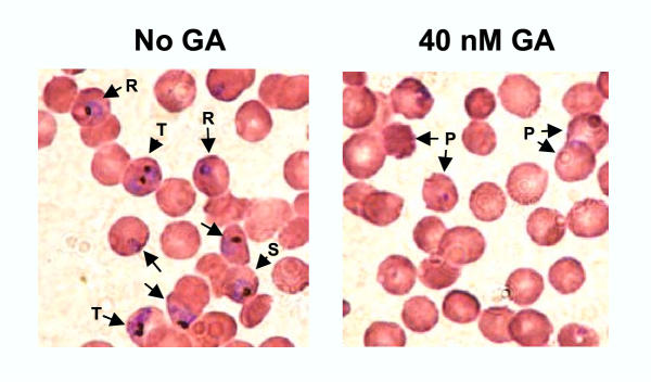
Figure 6.
Morphological changes in GA-treated P. falciparum. Asynchronous P. falciparum 3D7 culture was exposed to a range of GA concentrations for 48 h, and then thin smears of the cultures were made and stained with Giemsa. An equal volume of DMSO was added in the control cultures ("no GA"). A representative population exposed to 40 nM GA is shown. Infected erythrocytes containing stained parasites and a few stages are indicated with arrowheads (R = ring, T= trophozoite, S = schizont). Note the complete disappearance of parasites in the GA-treated culture and the appearance of pyknotic bodies (P).

