FIG. 4.
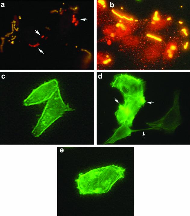
M1 protein induction of localized cytoskeleton rearrangement and stress fibers in HEp-2 cells is dependent on the PI 3-K pathway. (a and b) Wortmannin inhibited the ingestion of streptococci but did not impede adherence to HEp-2 cells. Infected cells were fixed, and the extracellular bacteria were stained with rabbit anti-GAS antibody followed by anti-rabbit-FITC conjugate. The cells were permeabilized, and intracellular bacteria were again exposed to rabbit anti-GAS antibody and labeled with anti-rabbit Cy3. Arrows indicate the intracellular bacteria, which appear red. Extracellular bacteria are yellow. (a) Cells infected in the absence of wortmannin. (b) Cells infected in the presence of 50 nM wortmannin. (c to d) Cells preincubated with purified M protein. (c) Control untreated cells. (d) Cells treated with 400 ng of recombinant M1 protein per ml for 10 min. (e) Cells treated with wortmannin plus 400 ng of purified M1 protein per ml in MEM with Fn. Arrows indicate actin tufts of lamellipodia and stress fibers.

