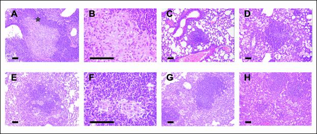
FIG. 4.
Evolution of granulomas in C57BL/6 (A through D) and DBA/2 (E through H) mice infected with isolate UTE 0423R at weeks 9 (A through C and E through G) and 22 (D and H) postinfection. (A, B, E, and F) Primary granulomas, with a center of infected macrophages and a mantle of lymphocytes. Panels B and F show intragranulomatous necrosis at a higher power (×400). The asterisk in panel A marks the zone amplified in panel B. (C and G) Secondary and tertiary granulomas, respectively, with a compact center of lymphocytes admixed with scanty infected macrophages. In panel G two secondary granulomas are surrounded by a layer of foamy macrophages that connects them both. (D and H) Tertiary granulomas, with septal thickening of alveoli filled with foamy macrophages. All microphotographs except those in panels B and E were taken at a magnification of ×100. Bars, 100 μm.

