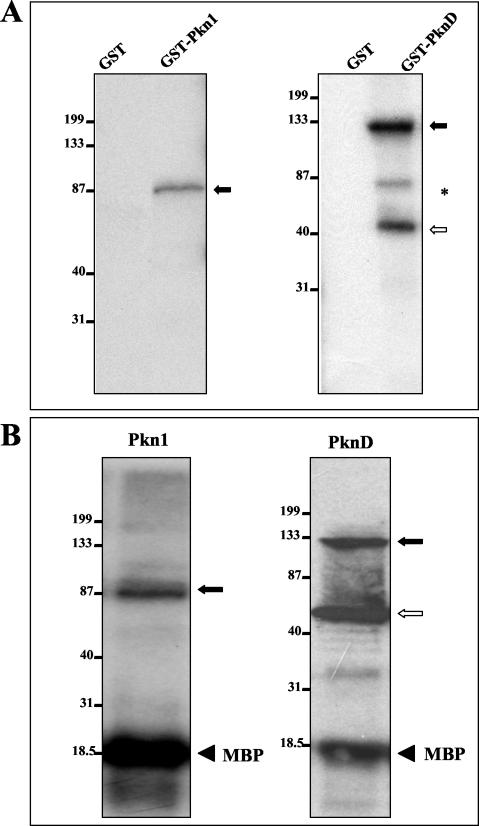
FIG. 3.
Autophosphorylation of E. coli-expressed GST-Pkn1 and GST-PknD. Purified GST-Pkn1 and GST-PknD fusion protein beads were directly used in the in vitro kinase assays. Kinase assays were performed as described in Materials and Methods, and proteins were separated by SDS-PAGE. After electrophoresis, the gel was transferred onto a nitrocellulose membrane and autoradiographed. (A) Solid arrows represent the expected phosphorylated products. The open arrow represents phosphorylation of the PknD protein fragment detected in the Coomassie blue-stained protein gel shown in Fig. 2A. *, a nonspecific phosphorylated product. Positions of molecular mass markers in each gel (in kilodaltons) are indicated on the left. In both gels, GST alone was used as a control. (B) MBP phosphorylation mediated by Pkn1 and PknD in the in vitro kinase reaction. The solid and open arrows represent phosphorylated Pkn1 and PknD as in panel A. Solid arrowheads represent phosphorylated MBP.

