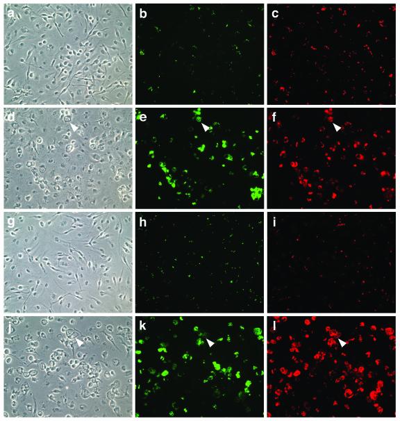
FIG. 1.
Analysis of Y. pestis and Y. pseudotuberculosis replication in macrophages by microscopy. C57BL/6 macrophages were infected with KIM10+/GFP (a to f) or IP2790c/GFP (g to l) at an MOI of 5. The infected cells were incubated for 4 h (a to c and g to i) or 25 h (d to f and j to l) and then fixed, and bacteria were labeled with a rabbit anti-Yersinia antibody (red). One hour before fixation, IPTG was added to the cells to induce GFP expression in viable bacteria (green). The samples were visualized by phase (left panels) or epifluorescence (middle and right panels) microscopy. Representative images were captured using a digital camera. Macrophages containing large numbers of intracellular bacteria displayed an enlarged and vacuolated morphology under phase-contrast microscopy (arrowheads).

