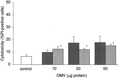
FIG. 3.
Effect of H. pylori OMV on gastric epithelial cell viability. AGS cells were grown alone or in the presence of 10, 20, or 50 μg of H. pylori OMV from a cag PAI+ toxigenic strain (dark gray bars) or a cag PAI− nontoxigenic strain (light gray bars). Cell viability was assessed by propidium iodide (PI) staining and flow cytometry after 24 h. Cytotoxicity is expressed as the percentage of propidium iodide-positive cells in each population. The data are means ± standard errors for five independent experiments. An asterisk indicates that the results for a treatment are statistically significantly different (P < 0.05) from the results for untreated cells.

