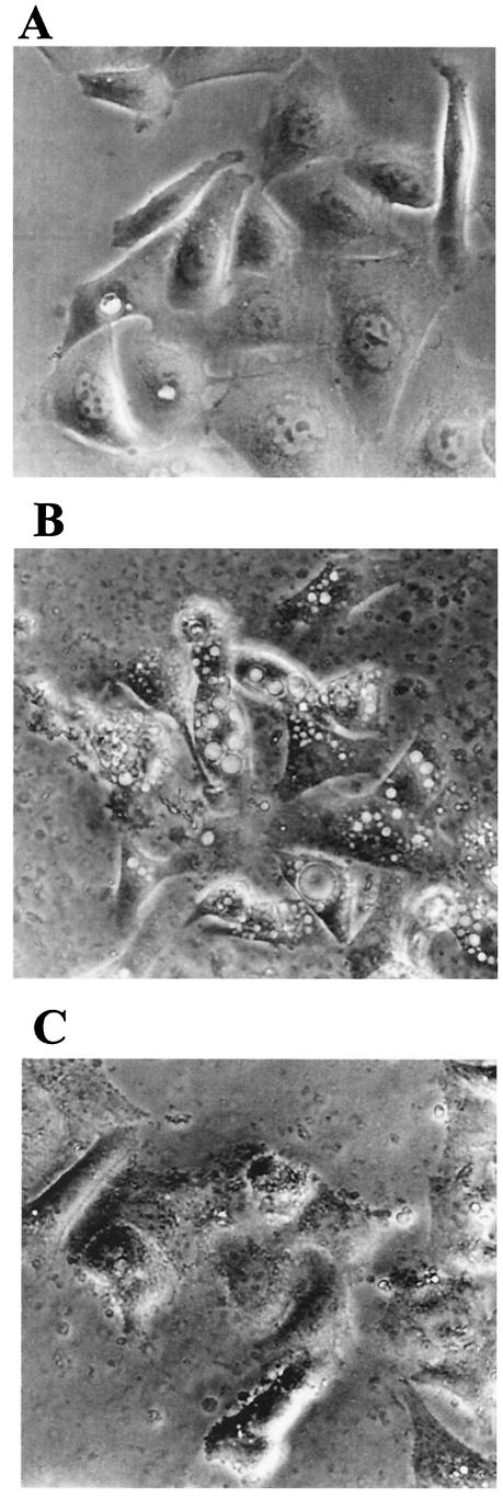
FIG.4.
Vacuolation in gastric epithelial cells treated with OMV. AGS cells were grown alone (A) or in the presence of H. pylori OMV (50 μg of protein) from a cag PAI+ toxigenic strain (B) or a cag PAI− nontoxigenic strain (C). Cell vacuolation was assessed by light microscopy after 24 h. The photographs are from a representative experiment.

