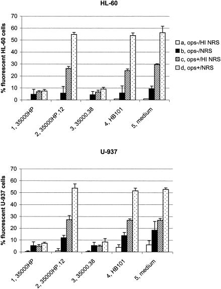
FIG. 3.
Uptake of opsonized and nonopsonized microspheres by HL-60 and U-937 cells exposed to H. ducreyi. HL-60 granulocytes (upper panel) and U-937 macrophages (lower panel) were incubated for 1 h at 33°C with H. ducreyi strains, E. coli HB101, or medium only (RPMI-F) followed by 1 h of incubation at 37°C with the following: nonopsonized microspheres in the presence of heat-inactivated NRS (a); nonopsonized microspheres in the presence of NRS (b); opsonized microspheres in the presence of heat-inactivated NRS (c); or opsonized microspheres in the presence of NRS (d). The percentage of cells containing intracellular fluorescent microspheres is shown. Each bar represents the mean with standard error from three independent experiments. 1, wild-type 35000HP; 2, lspA1 lspA2 double mutant 35000HP.12; 3, cdtABC hhdAB deletion mutant 35000.38; 4, E. coli HB101; 5, medium control.

