Figure 2.

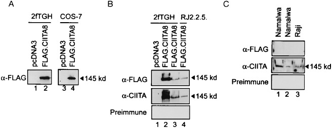
Detection of recombinant and endogenous CIITA. (A) Detection of transfected FLAG.CIITA8 by immunoblot. 2fTGH and COS-7 cells were transfected with 10 μg and 2 μg, respectively, of either FLAG.CIITA8 (lanes 2 and 4) or empty vector pcDNA3 (lanes 1 and 3). The cells were harvested 24 h after the transfection, and total lysates were prepared and separated on a 8% SDS/polyacrylamide gel. The gel was transferred onto nitrocellulose and the presence of recombinant CIITA was examined with 10 μg/ml α-FLAG antibody. (B) Transfected FLAG.CIITA8 was detected by both α-FLAG and α-CIITA antibodies. 2fTGH and RJ2.2.5 cells were transfected with either 10 μg (lanes 2 and 4) or 5 μg (lane 3) of FLAG.CIITA8 or control vector pcDNA3 (lane 1). Total lysates were prepared and examined with either α-FLAG antibody (Top), α-CIITA antibody (Middle), or preimmune serum (Bottom). (C) Anti-CIITA antibody specifically recognizes an endogenous CIITA, with an apparent molecular weight of 145 kDa. Two Namalwa and one Raji nuclear extracts were prepared and blotted with either the α-FLAG antibody (Top), α-CIITA antibody (Middle), or preimmune serum (Bottom).
