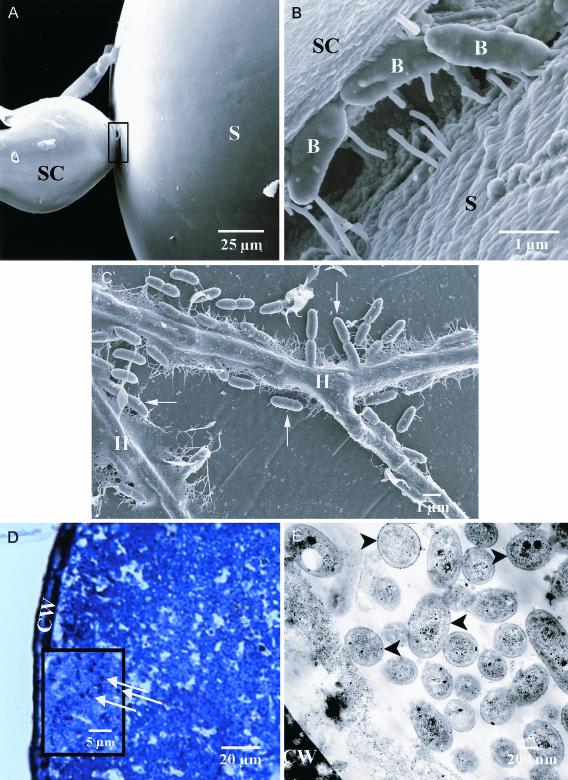
FIG. 2.
(A) SEM of a G. decipiens spore (S) with attached sporogenous cell (SC) inoculated with B. pseudomallei. (B) Field emission SEM of the junction between the spore and sporogenous cell shown in panel A. B, bacterium. (C) Field emission SEM of G. decipiens hyphae, adherent B. pseudomallei (arrows), and associated fibrillar material. H, hypha. (D) Optical semithin section of B. vietnamiensis-inoculated G. decipiens stained with toluidine blue. Bacteria (arrows) are present throughout the cytoplasm. CW, cell wall. (E) TEM of G. decipiens cytoplasm containing bacteria (arrowheads).

