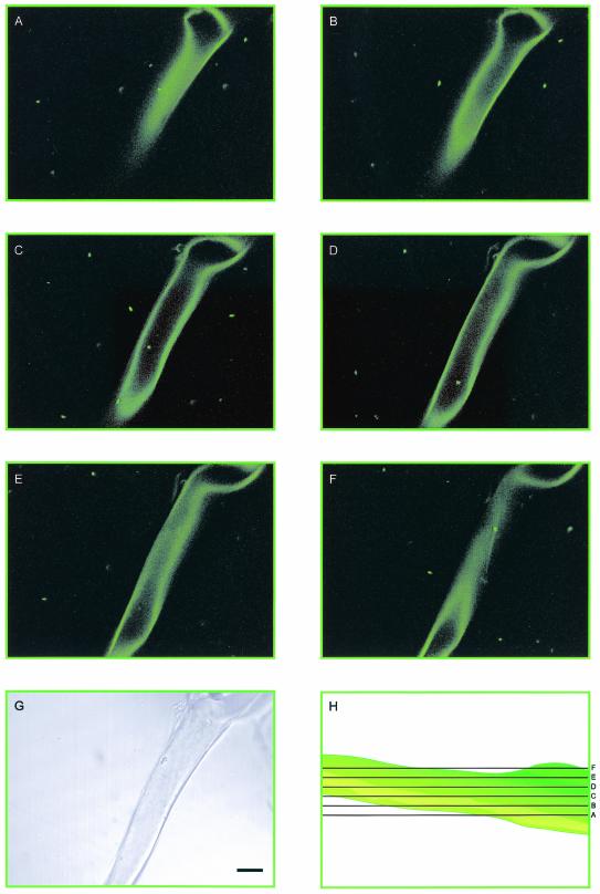
FIG. 3.
(A to F) Series of confocal laser scanning micrographs of GFP-labeled B. pseudomallei and a G. decipiens hypha. Motile, fluorescent bacteria are apparent in the aqueous medium, attached to the external surfaces of the hypha, and internalized within the hypha (C and D). (G) Nonconfocal transmission image. (H) Diagrammatic representation of the optical sectioning of the hypha. Bar, 30 μm. Images were falsely colored green using Adobe Photoshop 7.0.

