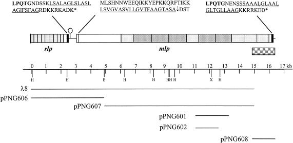
FIG. 1.
The 16,401-bp DNA region, showing the arrangement of mlp and rlp, and the λ and plasmid clones used to determine the sequence. The letters above the boxes indicate the cell wall sorting (black boxes) and secretion signals of the encoded proteins. The underlined sequences indicate hydrophobic domains. The shaded boxes indicate the encoded amino acid repeat regions, with light and dark boxes indicating the different-sized repeats in Mlp. The lollipop indicates a potential stem-loop structure. The checkered box indicates the DNA region which was amplified by PCR to determine the 3′ end sequence of mlp and to identify multiple mlp genes. The oligonucleotides Mlp-Paw-C and Mlp-C-check (Table 1) were used to amplify this region.

