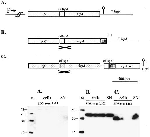
FIG. 3.
Gene constructs used to integrate into the L. fermentum BR11 bspA locus (top panels) and Western blot detection of fusion proteins in cell extracts and in the supernatant with anti-His5 antibody (bottom panels) for the native bspA genomic locus (A), the BspA-His6-CFTR fusion protein (B), and the His6-CFTR-Rlp fusion protein (C). In the diagrams in the top panels, the bspA upstream promoter (P→), the bspA terminator (T bspA), the rlp terminator (T rlp), and DNA encoding the BspA secretion signal (ssBspA), His6 (grey box), three copies of the CFTR peptide (hatched box), and the cell wall sorting signal of Rlp (rlp-CWS) are indicated. The DNA region which is the site of single-crossover homologous recombination into the bspA locus is stippled and marked with a cross. In the bottom panels, the Western blots contained His6 molecular mass markers (with sizes in kilodaltons) (lanes M); cell extracts prepared by boiling in 2× loading dye (lanes SDS), by sonication (lanes son), and with 5 M LiCl (lanes LiCl); and precipitated supernatant fractions (lanes SN). The amounts of cells or medium loaded in each lane are equivalent to 500 μl (lanes SDS), 50 μl (lanes son), 160 μl (lanes LiCl), and 625 μl (lanes SN) of culture.

