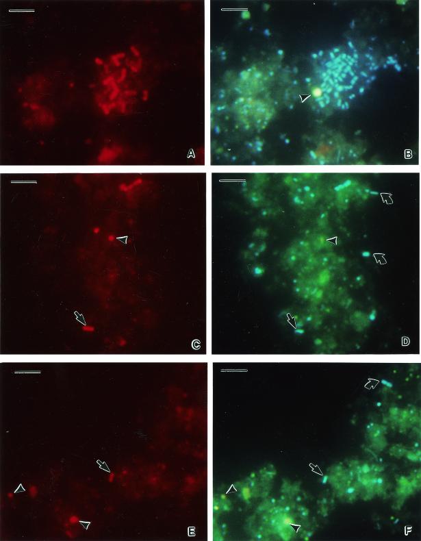
FIG. 2.
Whole-cell hybridization of cells extracted from T. testudinum sediment. (A) Hybridization with eubacterial probe EUB338; (B) identical microscopic field for DAPI staining; (C and E) hybridization with the C. orbicularis symbiont-specific probe Symco2; (D and F) identical microscopic fields for DAPI staining. Arrowheads, nonbacterial sediment particles; straight arrows, free-living form of C. orbicularis gill endosymbiont; curved arrows, environmental bacteria. Bars, 5 μm.

