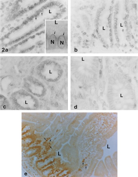
Figure 2.
HLA-H protein immunostaining in small intestine and colon. Distinct perinuclear signals are seen in the absorptive epithelium of duodenum (arrows in a and Inset). The Inset in a is a higher magnification photo showing staining around nuclei (N). Similar perinuclear staining is seen in jejunum (b and e) and ileum (c). In all of these segments, the reaction is localized to the intestinal crypts. This is seen most clearly in a lower magnification view of jejunum (e). In ascending colon (d), the signal is weaker and limited to the basolateral plasma membrane of the epithelial cells. Subepithelial leukocytes also show positive immunoreaction (see arrows in e). L, lumen. (a–d, ×400; a Inset, ×800; e, ×200.)

