
Fig. 4.
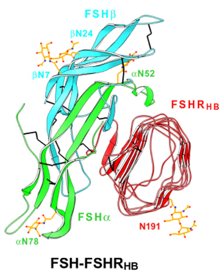
Ribbon diagram of the crystal structure of human FSH bound to FSHRHB. FSH α- and β-chains are in green and cyan, respectively. FSHRHB is in red. The observed N-linked carbohydrates at Asn52, Asn78 of FSHα , Asn7 and Asn24 of FSHβ , and Asn191 of FSHRHB are in yellow. Disulfide bonds are in black. Adapted with permission from Nature (Fan and Hendrickson, 2005a).