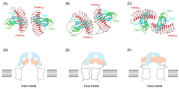
Fig. 5.

Dimeric associations. (A–C) Ribbon diagrams of quasi-symmetric crystallographic dimers of FSH-FSHRHB complexes, colored as in Fig. 4. The view is parallel to the symmetry axis, as if looking at the membrane. (D–E) Schematic illustrations of dimeric receptors on the cell membrane. Projected outlines from the complexes shown above and from rhodopsin are shown for the hormone (cyan), hormone-binding domain (orange), linker and 7TM transmembrane domain (black dotted trace). The view is perpendicular to figures directly above, parallel with the membrane plane. (A) Principal crystallographic dimer. (B) Principal dimer with outline of an offset 7TM dimer disposed for interaction with one of the bound FSH molecules. (C) Alternative crystallographic dimer. (D) Symmetric principal dimer with both 7TM domains engaged. (E) Asymmetric principal dimer with only one 7TM domain engaged. Note that dimer axes for the two components are not aligned. (F) Symmetric alternative dimer with both 7TM domains engaged.
