
Fig. 4.
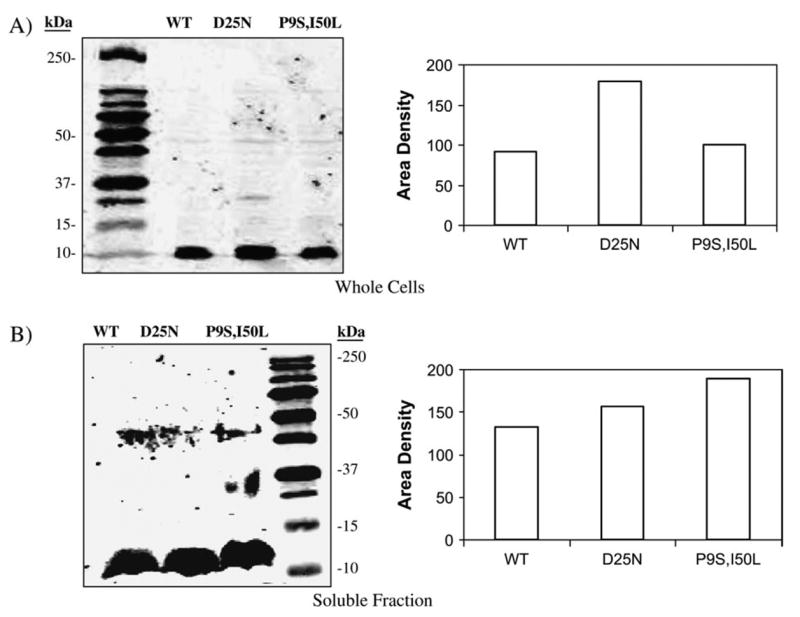
Expression levels of HIV PRs. E. coli transformed with WT, D25N or P9S/I50L HIV PR expression vectors were propagated to mid logarithmic phase in LB-ampicillin media. Gene expression was induced with 1 mM IPTG for 30 minutes. A) Whole cells were harvested by centrifugation, resuspended in 1 ml 1× SDS-PAGE running buffer, and analyzed by western blot analysis using an anti-HIV PR antibody. An IRD800 labeled secondary antibody and a LICOR Odyssey scanner were used to quantify the protein in each band. B) Cells were lysed, and the soluble fraction was removed and analyzed by western blot analysis.