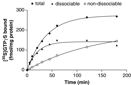
Figure 6.
[35S]GTPγS dissociation assays performed following different times of association. Membranes from CHO cells expressing the dopamine D2 receptor were incubated with 0.1 nM [35S]GTPγS in the presence and absence of 1 mM dopamine for different times and total bound [35S]GTPγS (total) was determined as described in the Materials and methods section. In parallel assays, [35S]GTPγS association was terminated at each time point by addition of 10 μM GTPγS and [35S]GTPγS dissociation determined after 30 min. Basal levels of [35S]GTPγS binding at each time point were subtracted from data. The figure shows the amount of [35S]GTPγS that was dissociable after 30 min at these different times (dissociable) and the amount that did not dissociate at these times (non-dissociable). Data shown are representative curves from an experiment performed independently three times with similar results.

