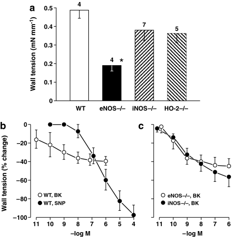
Figure 1.
Isolated ductus arteriosus from fetal mouse. (a) Comparison of contractile responses to L-NAME (100 μM) in WT vs NOS- and HO-2-deleted preparations (n, above columns). Baseline wall tension (mN mm−1) before treatment was as follows: WT, 0.04±0.02; eNOS−/−, 0.23±0.06; iNOS−/−, 0.28±0.06; HO-2−/−, 0.02±0.02. A significant difference (see asterisk) was found between WT and eNOS−/− (P<0.05; ANOVA). eNOS−/− also differed from WT in presenting a non-sustained L-NAME contraction in some cases (n=2). (b and c) Concentration–response curves to bradykinin, respectively, in WT (n=5 for both groups) and NOS-deleted (n=6 for both groups) preparations precontracted with indomethacin (2.8 μM). Wall tension before bradykinin was as follows: (b) WT, 1.04±0.17 (before SNP, 0.73±0.11); and (c) eNOS−/−, 1.05±0.08; iNOS−/−, 0.71±0.11. Progression of concentration-dependent changes is significant with each condition (P<0.001), whereas bradykinin relaxation is not different among genotypes (ANOVA in all cases). Where necessary, points have been offset to improve visibility, and any missing s.e. bar is within the size of the symbol.

