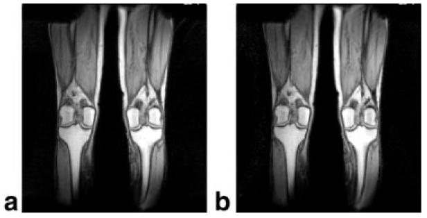
FIG. 10.

SENSE MRI of a normal volunteer acquired with four-element LPSA. a: Phased-array coronal image of a leg through the knees (TR = 150 ms, TE = 3.3 ms, NEX = 1, flip angle = 70, FOV = 48 cm, slice thickness = 7 mm, data matrix = 256 × 160, scan time = 25 s). b: Same section acquired with the same parameters, but for a reduction factor of 2 (scan time = 13 s).
