
FIG. 11.
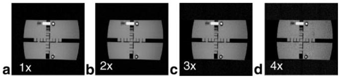
Images of the 27-cm-high, 20-cm-diameter phantom obtained with the GEM parallel reconstruction scheme. a: Fully gradient phase-encoded image. b–d: Images acquired with a reduction factor of 2 (b), 3 (c), and 4 (d). with a gradient-echo pulse sequence (TE = 6.7 msp, TR = 150 ms, flip angle = 30°, data acquisition matrix used to extract the sensitivity profile = 256 × 160, 256 × 256 points, NEX = 1, FOV = 34 cm, slice thickness = 5 mm). The coronal slice was 6 cm above the LPSA.