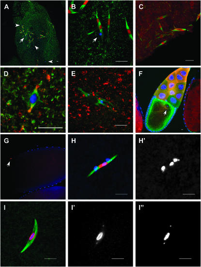
Figure 5.—
Subito lacking the N-terminal domain promotes formation of ectopic spindles. In all the experiments discussed below, the transgenes were expressed using the P{GAL4∷VP16-nos.UTR}MVD1 driver. DNA is blue; Subito-GFP, TACC (D and E), or Aurora B (C) is red, and tubulin is green. (A) Stage 14 oocyte expressing Subito with a deletion of the N-terminal domain fused to GFP (P{UASP:GFP-subΔNT}). The locations of chromosome masses are shown by the arrow and arrowheads. While these images are taken from oocytes also expressing wild-type Subito protein, similar effects were observed in oocytes lacking wild-type Subito protein. (B) The same oocyte as shown in A with the region around the arrow magnified. (C) GFP-subΔNT stage 14 oocyte stained for Aurora B (red). (D) In wild-type metaphase TACC (red) is visible at the poles. (E) In GFP-subΔNT ectopic spindles TACC (red) is absent. (F) In GFP-subΔNT stage 10 oocytes, there are no ectopic spindles. The oocyte karyosome is shown by an arrow. The cortical green staining is from microtubule growth that is nucleated from many organizing centers along the cortex of the Drosophila oocyte prior to NEB as part of the axis specification during oogenesis (Theurkauf et al. 1992; Cha et al. 2002; Steinhauer and Kalderon 2006). (G) Early GFP-subΔNT stage 14 oocyte with only one spindle, as indicated by a single patch of Subito staining (arrow). In the method used to prepare this oocyte, tubulin staining was not possible (see materials and methods). (H) A different GFP-subΔNT early stage 14 oocyte than in G, showing the premature separation of the karyosome. This was the only spindle in the oocyte. (H′) DNA channel, showing the separated small fourth chromosomes as well as the three remaining masses that are probably bivalents held together by chiasma. (I) Wild-type stage 14 oocyte stained for RCC1 (red). Separate channels for RCC1 (I′) and DNA (I″) show that even the small fourth chromosomes stain with RCC1. Bars, 10 μm.

