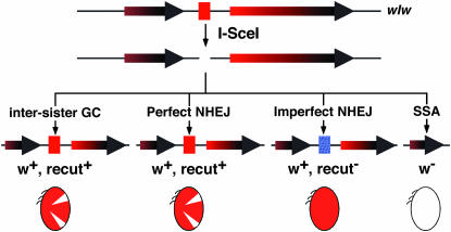
Figure 1.—
The hemizygous assay. The wIw insertion has two w genes: the copy to the left of the I-SceI cut site (red box) is nonfunctional (shorter arrow), and the copy to the right is a functional mini-w (longer arrow). The shading helps illustrate the part of w that is repeated. Four possible repair mechanisms are given below the I-SceI-generated DSB (middle), with the names of the mechanism on top and the phenotypic classifications at the bottom of the diagrams that depict the molecular structures of the different repair products. The blue box represents a mutated I-SceI cut site due to imperfect NHEJ. The ovals represent eyes with different degrees of pigmentation. A mosaic eye has both white and red areas.

