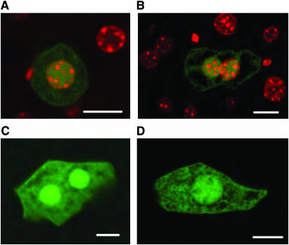
Figure 1.—
Green fluorescent mutant cells in the liver. (A and B) Merged images of EGFP signals and DAPI signals of mutant cells detected in the liver. The bright green fluorescent signals within these cells are colocalized with the DAPI-stained nuclei. (C and D) Images of EGFP signals of mutant hepatocytes. Bars, 10 μm.

