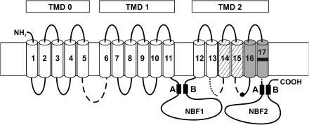
Figure 1.
SUR and the binding sites for openers and sulphonylureas. The transmembrane topology of SUR is according to Tusnády et al. (1997) and shows the organization of the 17 transmembrane segments in three TMDs, TMD0–TMD2. In the two nucleotide binding folds, the Walker A and B motifs are indicated. The binding site for the benzopyran and cyanoguanidine openers is formed by segments 16 and 17 and by a part (dotted) of the cytosolic loop linking segments 13 and 14 (Uhde et al., 1999). The black mark in segment 17 gives the approximate location of M1290 (SUR1) or T1253 (SUR2). The binding site of glibenclamide lies in segments 14 and 15 (hatched) and parts of the associated cytosolic loops, and in the loop linking TMD0 and TMD1 (broken lines). The black dot in the middle of the loop linking segments 15 and 16 represents S1237/Y1206 in SUR1/SUR2, which is of great importance for glibenclamide binding (Ashfield et al., 1999).

