Figure 8.
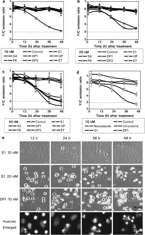
Screening results of S1 family compounds. HeLa-C3 cells grown in 96-well plates were treated with six podophyllotoxin family compounds plus ET at 10 nM (a), 20 nM (b) and 40 nM (c) for up to 48 h. Changes in the Y/C emission ratio were measured and used to compare the efficacy of each compound in inducing caspase-3 dependent apoptosis. (d) Apoptotic effects of DP1 and S1 were directly compared with the microtubule interfering agents nocodazole and vincristine, each at a concentration of 10 nM. (e) HeLa cells were treated with 10 and 20 nM S1 and 10 nM DP1 for up to 48 h. Phase images showing mitotic arrested cells with rounded cell morphology and apoptotic cells with cell shrinkage. Cells treated with 10 nM DP1 were also stained with Hoechst 33342, a DNA dye. The fluorescent images in the enlarged pictures show that 10 nM DP1 arrested cells in prometaphase at 12 h and caused DNA fragmentation after 24 h of drug treatment. The scale bars in the pictures of phase contrast and Hoechst staining are 50 and 25 μm, respectively. The error bars represent s.d. from three independent experiments.

