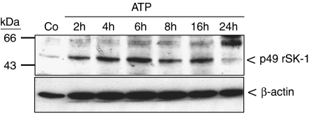
Figure 3.
Effect of ATP and UTP on SK-1 protein expression in renal mesangial cells. Quiescent mesangial cells were stimulated for 20 h with either vehicle (Co) or with 100 μM of either ATP for the indicated time periods (in h). Thereafter, cell lysates containing 100 μg of protein were separated on SDS-PAGE, transferred to nitrocellulose and subjected to a Western blot analysis using antibodies against either SK-1 (upper panel) or β-actin (lower panel) at dilutions of 1:1500 and 1:10 000, respectively. Bands were visualized by the enhanced chemiluminescences method according to the manufacturer's instructions. Data are representative of six independent experiments giving similar results.

