Figure 5.
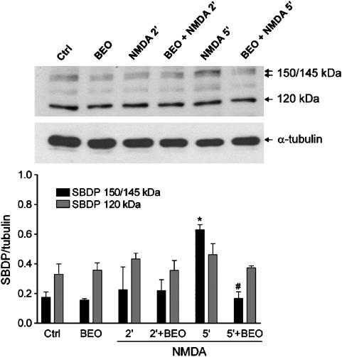
BEO prevented NMDA-induced activation of calpain I. Exposure of SH-SY5Y cells to 1 mM NMDA induced activation of calpain as assessed by Western-blotting analysis of generation of α-spectrin cleavage fragments (150/145 kDa) characteristic of calpain-mediated proteolysis. A significant accumulation of calpain-specific 150/145 kDa SBDP was reached at 5 min after NMDA exposure and this was prevented by a pretreatment (60 min beforehand) with BEO (0.01%). Exposure to NMDA and BEO, given alone or in combination, did not affect generation of 120 kDa α-spectrin fragment derived from caspase-mediated proteolysis. Histograms in lower panel show results of densitometric analysis of autoradiographic bands corresponding to 150/145 and 120 kDa SBDP. Data were normalized to the values yielded for α-tubulin. Each value is the mean±s.e.m. of three experiments. *P<0.05 vs control and #P<0.05 vs NMDA given alone (ANOVA followed by Tukey–Kramer multiple comparisons test).

