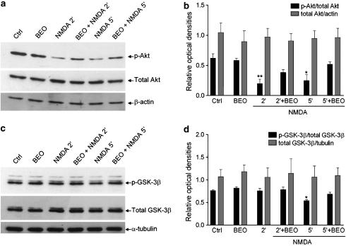
Figure 6.
BEO reduced NMDA-induced decrease of phospho-Akt and phospho-GSK-3β levels in SH-SY5Y cells. (a). Exposure of SH-SY5Y cells to 1 mM NMDA for 2 and 5 min induced deactivation of Akt kinase as determined by Western-blot analysis of phospho(Ser473)-Akt (p-Akt) levels and this was attenuated by BEO (0.01%) applied to neuroblastoma cultures 60 min before NMDA exposure. (b) Densitometric analysis of immunoreactive bands shows that the significant changes in phospho(Ser473)-Akt levels induced by BEO in NMDA-treated cells were not associated with changes in total Akt immunoreactivity. (c) Exposure of SH-SY5Y cells to 1 mM NMDA for 5 but not for 2 min induced activation of GSK-3β kinase as determined by Western-blot analysis of phospho(Ser9)-GSK-3β (p-GSK-3β) levels and this is attenuated by BEO (0.01%) applied to neuroblastoma cultures 60 min before NMDA addition. As confirmed by densitometric analysis of immunoreactive bands (d), BEO did not affect the expression of total GSK-3β in NMDA-treated cells. Each value in b and d is the mean±s.e.m. of 3–6 experiments; *P<0.05 and **P<0.01 vs control, respectively (ANOVA followed by Tukey–Kramer multiple comparisons test).

