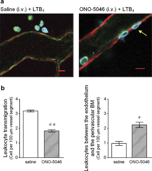
Figure 6.
Analysis of the site of arrest of leukocytes in mice treated with the neutrophil elastase (NE) inhibitor. Cremaster muscles from wild-type (WT) mice treated with intravenous saline (control) or with the NE inhibitor ONO-5046 (see Materials and methods for dosing regime) and stimulated with topical LTB4 (10−7 M) were dissected away from mice for immunostaining and analysis by confocal microscopy. Briefly, tissues were fixed in paraformaldehyde and immunostained overnight by using primary antibodies directed against mouse laminin 10 (as a marker for the endothelial cell basement membrane; red), PECAM-1 (as a marker for endothelial cells; green) and MRP14 (as a marker for neutrophils; blue), as detailed in Materials and methods. Samples were analysed by confocal microscopy as detailed in Materials and methods. (a) The figure shows representative images of postcapillary venules from saline-treated (left) or ONO-5046-treated mice (right) captured by confocal microscopy. While the image from the saline-treated mouse shows several leukocytes in the extravascular tissue, the tissue from ONO-5046-treated mouse shows clear evidence of leukocytes trapped within the vessel wall. (b) The graphs show quantification of leukocytes transmigrated into the surrounding tissue (left-hand graph) or trapped within the vessel wall (right-hand graph), as observed by confocal microscopy. Leukocyte transmigration (left panel) was quantified as number of cells in the tissue within 50 μm of the vessel wall across a 100 μm vessel segment. Within the same vessel segments, the number of leukocytes trapped between the basement membrane and the endothelial cells (right panel), following LTB4 stimulation (60 min), was quantified. Results represent a mean of 5–8 tissues analysed and a significant difference between saline- and ONO-5046-treated groups is indicated by asterisks; *P<0.05 and **P<0.01. (For colour figure see online version.)

