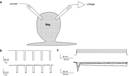
Figure 1.
Electrophysiological techniques. (a) Diagram of the location of two micropipettes for making current–clamp and voltage–clamp recordings from bag region of Ascaris suum somatic muscle. (b) Current–clamp recording showing membrane potential change (lower trace) in response to 0.5 s, 40 nA, current pulses (upper trace). Input conductance 3.7 μS. (c) Voltage–clamp recording showing current responses (leak subtracted lower trace) in response to depolarising potentials from a holding potential of −35 mV. Note the presence of the voltage-activated transient inward current and a sustained current. Sustained currents were not prominent in all cells.

