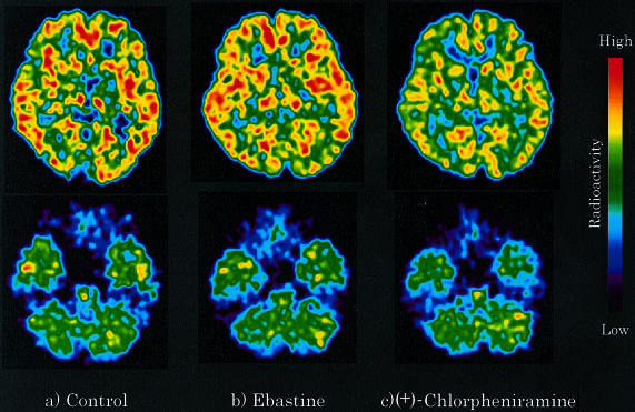
Figure 1.
Brain distribution of [11C]-doxepin radioactivity was examined in healthy male subjects by PET after the treatments of antihistamines. (a) Control (nondrug treatment), (b) ebastine 10 mg treatment, and (c) (+)-chlorpheniramine 2 mg treatment. Typical representatives of PET images are shown at the striatal and cerebellar levels. The images were obtained at 45–90 min after the injection of [11C]-doxepin.

