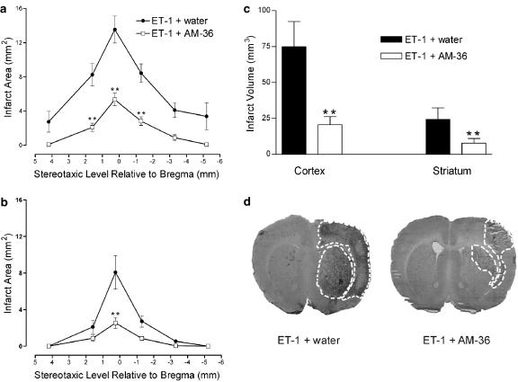
Figure 3.
Effect of AM-36 on infarct area (a, b) and total infarct volume (c) in both the cortex (a) and striatum (b) induced by ET-1, with data presented as mean±s.e.m. of infarct area measured at six predetermined coronal planes through the brain. **P<0.01 (a, b) compared with vehicle-treated control rats (Two way repeated measures ANOVA followed by Student–Newman–Keuls method) for infarct area. **P<0.01 (c) compared with vehicle-treated control rats (Student's t-test). Infarct area and volume is significantly reduced following AM-36 treatment. Representative images generated from unstained sections, using the MCID, showing reduced infarct size from vehicle-treated (ET-1+water) rats following treatment with AM-36 (d). Infarct area in right hemisphere induced by ET-1 can be visualized as the dark area and is marked by the white dotted line on each of the sections. n=5–6 rats in each treatment group.

