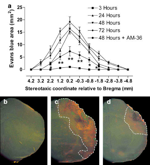
Figure 4.
Time course of Evans Blue extravasation in rat brain from 3 to 72 h after ET-1 induced MCAo (a) including the effect of AM-36 when administered up to 48 h, with data presented as mean±s.e.m. of area stained by Evans Blue. The area stained by Evans Blue significantly increased from 3 to 48 h, and then decreased at 72 h (P<0.001, 3 versus 24 h; P<0.05, 24 versus 48 h; P<0.05, 48 versus 72 h). *P<0.05, **P<0.01 AM-36 compared to ET-1 group at 48 h (Two way repeated measures ANOVA followed by Student–Newman–Keuls method). n=6 rats in each group. Representative sections through the ipsilateral cortex ( × 2) viewed following Evans Blue injection at 48 h (b–d), showing reduced Evans Blue extravasation with AM-36 administration (d) when compared with vehicle-treated rats (c). There was no extravasation of Evans Blue in the sham-MCAo rats (b). Area of Evans Blue extravasation is indicated by white dotted line.

