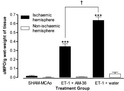
Figure 5.
MPO activity was measured in the ischaemic and non-ischaemic hemispheres of ET-1+ water, ET-1+AM-36 and sham-MCAo groups. Values are mean±s.e.m., expressed as units of MPO activity per gram wet-weight of brain tissue, for n=5–6 rats in each group. AM-36 treatment significantly reduced the MPO activity in the ischaemic hemisphere when compared to the ET-1+water group. ***P<0.0001 ischaemic hemisphere versus non-ischaemic hemisphere for ET-1+AM-36 and ET-1+water groups; †P<0.05 AM-36 treatment group compared with the vehicle-treated group (ANOVA followed by Student–Newman–Keuls method).

