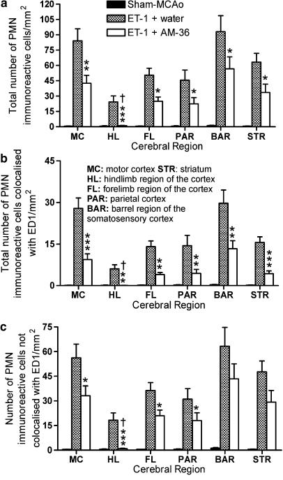
Figure 6.
Cell counts of polymorphonuclear neutrophil (PMN) anti-sera and ED1 immunoreactive (IR) cells following double staining at 48 h after ET-1 induced middle cerebral artery occlusion (MCAo) in selected cerebral regions. PMN IR cells were increased at 48 h after MCAo. (a) Total number of PMN IR cells at 48 h following MCAo in different cerebral cortex regions. AM-36 treatment significantly reduced the total number of PMN IR cells in all of the regions investigated when compared to vehicle-treated rats. AM-36-treated rats were significantly different from sham-occluded rats in all regions except the hindlimb region of the cortex. (b) The total number of PMN IR cells that were colocalized with ED1. AM-36 administration reduced the total number of PMN IR cells that were colocalized with ED1 in all cerebral regions examined, except the hindlimb region of the cortex, which again showed no difference from sham-occluded rats. (c) The total number of PMN IR cells not colocalized with ED1 was also reduced by AM-36, however, there was no significant difference between ET-1 and ET-1+AM-36 treatment in the barrel region of the somatosensory cortex and the striatum. Bars are presented as mean±s.e.m. of the number of IR cells in each cerebral region. *P<0.05, **P<0.01, *** P<0.001 AM-36 compared with vehicle-treated rats; †P>0.05 AM-36 compared with sham-occluded control rats (Kruskal–Wallis ANOVA with Dunn's post-test). n=6 for both ET-1+water and ET-1+AM-36-treated rats and n=5 for sham-occluded control rats.

