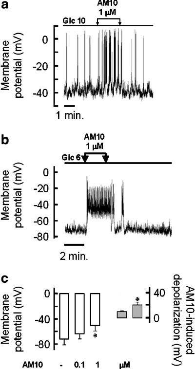
Figure 6.
Effects of AM10 on the native pancreatic β cell membrane potential. (a and b) The membrane potential of a single mouse β cell was monitored in current-clamp mode of the patch-clamp technique. The glucose concentration of the perfusion medium was maintained stable (10 and 6 mM, in (a) and (b), respectively) and AM10 (1 μM) was added and withdrawn as indicated by the arrows. (c, left part) Average of the membrane potential of single β cells perfused with a solution containing 6 mM glucose, in the presence or not of AM10 at the indicated concentration. (c, right part) Average of the AM10 induced depolarization (0.1 and 1 μM left and right bar, respectively), *P<0.05 vs CT. (a) and (b) Traces representative of three and five experiments, respectively. (c) Mean±s.e.mean of nine experiments (four and five for 0.1 and 1 μM of AM10, respectively).

