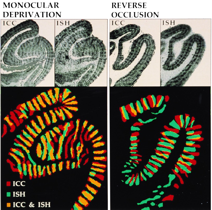
Figure 1.
Visualization of OD columns in coronal sections of area V1 by Zif268 mRNA and protein staining. (Upper) ICC and ISH staining results from adjacent sections. (×2.2.) (Lower) The OD columns were delineated, color-coded, and digitally superimposed. (×3.8.) The protein-positive and mRNA-positive regions show significant overlap in the MD condition, while they are largely complementary in the RO condition.

