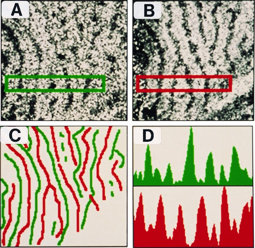
Figure 2.
Topographic organization of mRNA- and protein-stained ocular dominance columns in flattened views of area V1. (A) Film autoradiogram showing OD columns labeled by zif268 mRNA. (×7.2.) (B) Immunostained section showing OD columns labeled by Zif268 protein. (×7.2.) (C) Topographic layout of zif268 mRNA (green) and protein (red) labeled columns shows that they have a complementary, nonoverlapping arrangement. (×7.2.) (D) Optical density profiles confined to the rectangular region in the stained sections show that regions of intense mRNA labeling are accompanied by poor protein staining and vice versa.

