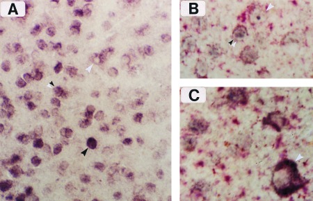
Figure 3.

Cellular resolution of zif268 mRNA and protein labeling by simultaneous ISH (dig-conjugated complementary RNA probe) and immunostaining. (A) Border region of two OD columns in layer II, showing single-labeled cells that are mRNA-positive (white arrowhead) or protein-positive (black arrowhead). Double-labeled cells are also visible in this figure (double arrowhead), and they are enhanced when differential color staining is applied, as in B and C. (×250.) (B) Immunoreactive product (blue) and dig-mRNA complex (red) can be seen together in several double-labeled cells from layer II. (×400.) (C) Meynert cell from layer VI (white arrowhead) enriched with dig-conjugated reaction product surrounding a negatively stained nucleus. (×400.)
