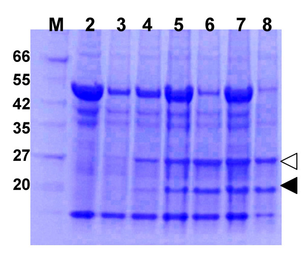
Figure 4.
Temporal analysis of GFP expression from TMV vectors. N. benthamiana leaves were infiltrated with an A. tumefaciens/pJL43:GFP cell suspension. Total soluble protein extracts were prepared from infiltrated leaf tissue from 3–7 days post infiltration (DPI) or from plant tissue systemically infected with the TMV:GFP vector (12 DPI). Extracts were separated on a 4–20% SDS PAGE gel and stained with coomassie blue. Lanes: M, MW marker; 2, Healthy leaf extract; 3–7, extracts from A. tumefaciens/pJL43:GFP infiltrated leaves, 3–7 DPI, respectively; 8, extract from systemically infected tissue, 12 DPI. Marker band sizes (in kDa) are listed. Locations of GFP and TMV CP are identified by open and solid triangles, respectively.

