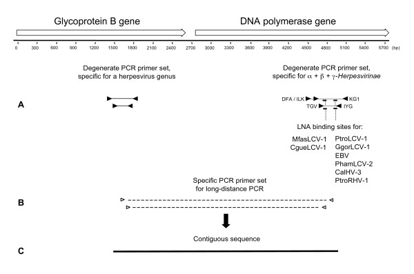
Figure 1.
LNA-based and bigenic amplification of beta- and gammaherpesviruses. Schematic diagram of the analysis strategy. (A, right) Initially, panherpes nested PCR with deg/dI primers (black triangles) is performed for amplification of DPOL sequences. In the first round, primers for amplification of 710 bp and 480 bp are used simultaneously, either in the absence or presence of LNA. The binding regions of the LNA are present in the amplified sequences of both the first and the second PCR round, and represented by short thick lines. The targeted viruses are indicated. (A, left) The binding regions of genus-specific deg/dI gB-primers are indicated by black triangles. Amplimers of the first and the second PCR round are 320 bp and 250 bp, respectively. (B) After both gB and DPOL sequences were determined, long-distance nested PCR (dashed lines) was performed with specific primers (open triangles) binding to gB (sense) and DPOL (antisense). (C) A final contiguous sequence of approximately 3.5 kbp was obtained (solid line).

